


Welcome to our patient resource page.
At SonoAid Imaging we are committed to providing the highest quality in care to our patients. We continuously refine the services we provide, monitor and measure the diagnostic procedures our patients receive and evaluate our performance against our own rigorous standards as well as industry benchmarks. We strive for excellence in the care we provide, and a part of that care is providing education and information for the physicians and patients about the services we offer.
What is Ultrasound?
 Ultrasound imaging, also called ultrasound scanning or sonography is a method of obtaining images from inside the human body through the use of high frequency sound waves. The device, called a transducer, sends out high frequency sound waves and then “listens” to the response. The reflected sound wave echoes are recorded and displayed as a real-time visual image. Ultrasound is considered a safe, effective and painless procedure that can be used for a wide range of diagnostic purposes, as well as for assisting with minimally invasive procedures such as needle biopsy and needle aspiration, as it provides real-time imaging of the soft tissues of the body. There is no ionizing radiation or x-rays used during the ultrasound procedure, and it is generally easier to perform and less expensive for both patient and doctor than other imaging procedures. Ultrasound imaging is considered safe for all patients with no known risks. It can be repeated as often as needed. Ultrasound is a useful examination tool for many of the body’s internal organs, including the heart, arteries, veins, liver, gallbladder, spleen, pancreas, kidneys, and bladder. Because ultrasound images are captured in real-time, they can show movement of internal tissues and organs, and enable physicians to see blood flow and heart valve functions. Ultrasound can be used to produce images of the internal structures within nearly any area of the body, allowing for clear, detailed visualization of a wide range of conditions. Masses, tumors, infections and other abnormalities can be easily detected using ultrasound. This procedure can also be used as imaging guidance for biopsy, needle placement and other types of treatment.
What is the Doppler?
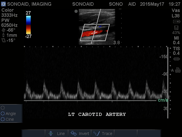
Doppler ultrasound is a special ultrasound technique that evaluates blood as it flows through a blood vessel, including the body’s major arteries and veins in the abdomen, arms, legs and neck.
There are three types of Doppler ultrasound:
Color Doppler uses a computer to convert Doppler measurements into an array of colors to visualize the speed and direction of blood flow through a blood vessel.
How is the ultrasound performed?
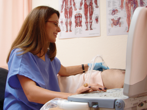 All ultrasound procedures are considered safe and painless, with no major side effects or complications. In most cases, the exam is performed by applying a gel to the skin, before moving a transducer back and forth to emit sound waves and create images of the organs, tissues, muscles, ligaments, vessels and other structures within the body. The transducer separates and identifies the different echoes which are changed into electrical energy by the ultrasound machine which shows up as images on the screen. The procedure begins with the patient lying down on the examination table as a water-based gel is applied to the area on their body that will be observed. This gel allows consistent contact between the body and the transducer, free of any air pockets that could get in the way. The transducer is kept firmly against the skin and is “swept” back and forth across the area to allow for the most detailed observation possible. A certified technologist will perform the exam. The whole process usually takes 30 minutes. There is no discomfort associated with this procedure; if the part of your body being observed has already been tender there will be some slight pressure against it. The results of your ultrasound procedure are analyzed by our board certified radiologists or cardiologists: physicians who specialize in the interpretation of imaging examinations. The results will then be sent to your primary or referring doctor. In some cases, our radiologists and cardiologists will discuss the outcome of your exam with your doctor upon completion of the procedure.
How should I prepare for the Ultrasound?
 being imaged, you may be asked to wear a gown during the procedure. Also, it is important to inform your doctor if you have taken a barium enema or had any other upper gastrointestinal tract tests within the past few days, as they can severely distort the ultrasound image. If any part of your gastrointestinal tract, aorta, liver or pancreas must be observed, then you will be advised to stop eating for the six hours prior to the exam. If your kidneys, urinary bladder or pelvic organs are the targets, in addition to the aforementioned eating requirements you may need to drink several glasses of water one hour prior to your exam and avoid urinating so that your bladder is full when the scan begins. Further instructions are listed on your referral sheet and will be clarified during the scheduling process. of the procedure.
Will my insurance cover these tests?
With our conveniently located office in Glendale, we can easily accommodate same day appointments for our patients who reside in Glendale, La Crescenta, Burbank, Eagle Rock, Pasadena and Los Angeles.
Please plan to arrive a few minutes ahead of your scheduled appointment. You may be asked to fill out paperwork. The following information needs to be available:
- Your photo ID.
- Your Insurance Card
- Your Physician’s order or prescription
- Your medical history. (For certain procedures)
At SonoAid Imaging our objective is to make your visit as comfortable and pleasant as possible. Our physicians and staff are dedicated to assisting you with your imaging needs and providing your doctor with quick and accurate results.
For more information on services and what to expect during your exam, please call as at (818) 241-2514. Our highly professional staff looks forward to assisting you.